
One of the first things you will learn in an Emergency Medical Technician program is a basic run down of what an assessment will look and sound like. I don’t just mean what you say to a patient and what to ask of them. The moment you are dispatched on a call your assessment should start. What are you being dispatched for? Where are you being dispatched to? How did the emergency happen? These questions will help you decide what to bring with you on scene, what scene hazards there may be, and you can start to figure out what kind of information you might need to gather from the patient and the scene. It might also help you determine how quickly you and your partner may need to work.
One of the most important things for you to be aware of is that if you are enrolled in a quality EMT program the skills and information you are going to learn all revolve around making sure you can pass the National Registry Psychomotor and Cognitive exams. It does not mean what you learn will have you ready to be fully operational in the field and most companies you will be interviewing with for a position will understand that. With that being said, the assessments you will learn are pretty consistent with how you will conduct them in the field. They are broken down by the type of emergency; trauma emergencies or medical emergencies. As suggested by the categories, depending on the nature of the emergency the assessment you will conduct with vary slightly which makes logical sense due to needing to know different information to treat the patient effectively. The scene size up, primary assessment, history taking, and reassessment sections are practically mirrored between the categories leaving the secondary assessment as the deviated section. In this post I will be addressing the scene size up and primary assessment portions of an assessment.
What I stated earlier in regards to thinking about how you were dispatched and where you are going, is not a part of the National Registry Psychomotor examination skill sheet. However, in doing so it will help you be prepared for when you get on scene. In accordance to the skill sheet the first thing you do after physically arriving to a scene is the scene size up. This section is important because it allows you to determine if you should approach the scene and if you do approach the scene what additional units do you need for support. Even though EMTs are by definition people who respond to emergency situations, it is very important that the safety of the EMS crew comes first. Therefore it is crucial that you do not develop tunnel vision and focus solely on the patient because if you end up injured in some way, now there are two people in need of emergency services and your partner becomes the sole provider on scene. You should also be cognizant of your skill set and the protocols in your area. EMTs are basic life support providers. If you arrive on scene and your patient clearly needs a treatment that is outside of your scope of practice, you should call for additional support. Dispatchers are never going to be 100% accurate since they can only rely on information given over the phone by the caller and you should be prepared to request additional resources when necessary.
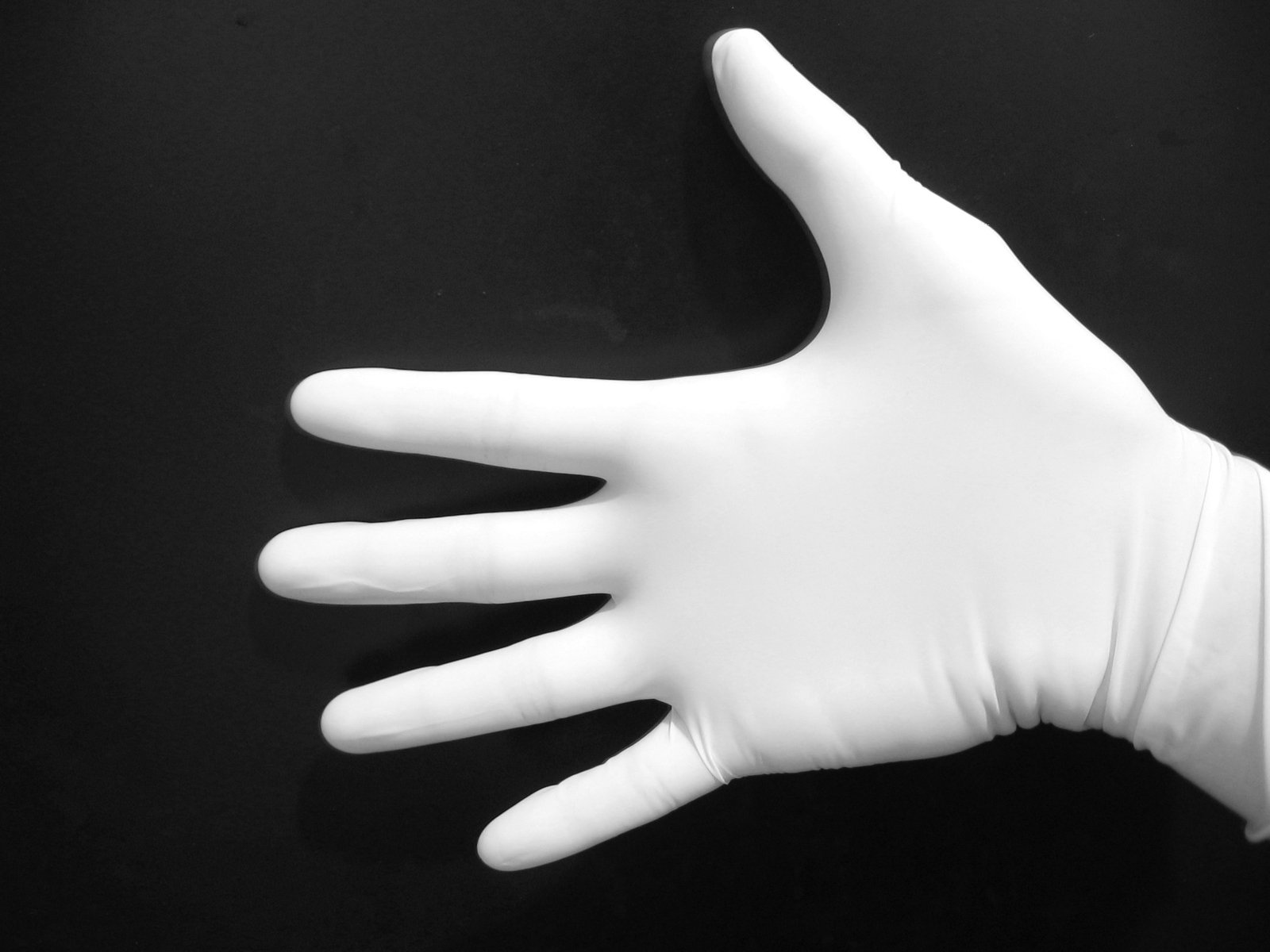
The other components of the scene size up include putting on the proper personal protective gear (gloves, gowns, etc.), determining the true cause of the emergency, and considering the need for spinal stabilization. Even though most season providers do not wear gloves on every call, I personally recommend doing so. You never know what you might put your hand in on scene or when the condition of the patient my change and I don’t want to take anything back with me. The patient or a family member may have coughed all over the table you put your hand on and now you’re putting your hand all over your cell phone and next thing you know you’re sick. So at the very least wear gloves on every call for your own health. Determining the cause of the emergency can be tricky some times, but it’s very important to get as much of the truth as possible so that when the patient is being treated in the emergency room at the hospital they don’t have an adverse reaction to a medication or you don’t cause more damage to an injury you might not have initially noticed. The nature of the illness or the mechanism of injury can also lead you down a line of questioning or cause you to notice to things you might have missed otherwise. Spinal stabilization is vital when it is needed. Due to the fragility of the spinal cord whenever there is even the potential for damage you should apply a cervical collar on a patient otherwise there may be long term irreversible consequences. The potential for such damage is something you can ascertain from the mechanism of injury.
After you determine it is safe for you and your partner to approach, you’ve protected yourself from bodily fluid contact, determined the possible cause of the emergency, called for any additional resources necessary, and considered whether or not the patient will need a cervical collar you can make initial patient contact. Even though that sounds like a lot, most of these steps are accomplished within a minute of arriving on scene. However just because you completed these objectives at the beginning of the call, it does not mean that you stop considering them. Emergency scenes are fluid environments and you should alway be considering your safety and the safety of your partner throughout the call. Remember, do not develop tunnel vision.
The second minute spent on scene will consist of your primary assessment. The primary assessment basically helps you determine how bad the emergency is. You will need to determine the patient’s level of consciousness using the AVPU scale. AVPU is an acronym that stands for Alert, Verbal, Pain, or Unconscious. A person who is alert will be conscious, responding to their environment, and probably reacting in a manner appropriate for their emergency (i.e. writhing in pain, groaning, etc.). A person that falls under the verbal category will respond when spoken to. Under the pain category the person will respond to a painful stimuli such as a pinch on the ear lobe or a sternal rub. A person who is unconscious will fail to respond to any stimuli. After that you will need to determine what the patient’s chief complaint is and if there are any apparent life threats. Most of the time this can be determined either by asking the patient or by simply looking at the patient. In emergencies it is possible for the patient to have multiple complaints, but it’s important to prioritize them to ensure the most positive outcome possible. It will also help you to figure out what you should do next when treating the patient.
After figuring out what caused you to be dispatched to the scene you need to evaluate the patient’s airway, breathing, and circulation (known as their ABCs throughout my EMT certification program) as part of the primary assessment. The patient’s airway is in reference to the physical airways structures such as the naso-, oro-, and laryngopharynx as well as the trachea, bronchi, bronchioles, and alveoli. The organs and muscles within the human body require a constant supply of oxygen and when they do not fulfill their oxygen needs, it can cause the cells of the deprived areas to die off. Sometimes rather quickly and irreversibly. So insuring that the patient’s airway is patent, or clear, will help you as the provider ensure the components of the body are getting the oxygen they need. The last thing you need is additional complications arising. When a patient’s airway is not patient, you will typically hear either a stridulous, wheezing, or snoring sound coming from the patient. To correct the airway obstruction, try to first align the head in a neutral, mid-line position and either perform a head tilt chin lift maneuver or a jaw thrust maneuver. These maneuvers prevent the tongue in the patient’s mouth from blocking the entrance to the oropharynx. If after realigning the head the patient continues to have an obstruction, you can either insert a naso- or oropharngeal airway device. The devices are inserted the patient’s nose and mouth respectively and assist in providing a clear passage for oxygen to travel into the lungs.
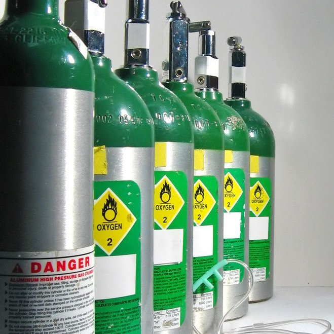
The next step will be to evaluate the patient’s breathing in reference to the actual act of breathing. You should take notice to the rate in which the patient is breathing and how much effort they are putting into breathing. Most adults at rest take between 12 and 20 breathes per minute. If a patient is taking less that 12 breathes per minute, they are likely not taking in enough oxygen to sustain themselves and it’s important that you either assist them with breathing or you supply supplemental oxygen to them. If a patient is taking more than 20 breaths per minute, they are likely taking in too much oxygen. That may seem strange given that the body’s cells require oxygen to function, so why would having an excess amount be considered a bad thing? Without diving into the pathophysiology behind it too deeply, the body functions best when it achieves homeostasis. Homeostasis is when the body reaches an equilibrium, meaning that there is just the right amount of bad and good things in the body. When a patient breaths at a rate that’s faster than normal, they are also subsequently breathing out more carbon dioxide. Without an appropriate level of carbon dioxide the blood vessels to the brain may constrict causing oxygen deprivation which can lead to a multitude of complications. Therefore slowing a patient’s breathing would be appropriate in this case.
When it comes to the amount of effort a patient is putting into breathing, you should look for additional muscle usage and listen to the types of breaths being taken. When a patient is using accessory muscles to breathe, you should see movement around the shoulders, neck, and upper chest when breathing in and movement around the abdomen when breathing out. You may also notice retractions, which is when the intercostal muscles (muscles between the ribs) sink into the rib cage when breathing in. These are all atypical muscle movements and indicate that the patient is putting additional effort into the act of breathing. If a patient continues to use accessory muscles to breathe, it can cause fatigue and the patient may become unable to breath on their own. Atypical breathing sounds include those mentioned earlier, stridulous, wheezing, and snoring, but you should also take into consideration the amount of force it sounds like each breath is using. For example when breathing normally, there is little to no sound noticeable, but think of what it sounds like after a run, or when a mother is in labor, or when someone sighs. You can hear the additional force used in each breath. This is referred to as labored breathing. Along with the increase in force there is an increase in the effort of the muscle to inhale/expel the air. As with accessory muscle usage, the longer a patient uses those muscles the more likely the muscles will become fatigued and cause additional problems.
Once you take the patient’s airway and breathing into consideration, you need to evaluate the patient’s circulation. To do so there is three major things to pay attention to; major bleeding, pulse, and skin. Major bleeding is pretty obvious in most cases in that if a patient is bleeding profusely externally you will see it. You can attempt to stop the bleeding as best you can with gauze, direct pressure, and tourniquets. However in considering the mechanism of injury during your scene size up, you should also be thinking about major bleeding internally. There is very little you can do as an EMT for major internal bleeding, but you can continue to monitor a patient’s blood pressure and give the hospital a report to have the appropriate staff on standby.
When considering the patient’s pulse in a primary assessment, you are not actually measuring the pulse. What you are feeling for is the presence, strength, and the rhythm of the pulse. Initially I do not care about the actual rate of the pulse; I need to figure out if one is present so I can consider initiating CPR, if it feels as though the heart is effectively moving blood throughout the body, and if the patient’s heart is functioning in a normal pattern. This information will help me determine the severity of the emergency so I know whether or not to call for additional support like a paramedic and so I know the likelihood of the patient’s affect deteriorating quickly.
In reference to the patient’s skin, you need to evaluate the temperature, color, and dryness. Abnormal temperature can indicate hyperthermia, hypothermia, infection, and fever among other things. This will allow you to consider other avenues of treatment for your patient and may even re-prioritize the complaints given by the patient. It’s not uncommon for some patient’s to be overwhelmed by how they feel that they cannot accurately describe everything that is wrong with them, especially children. The color of a patient’s skin can help you anticipate how the status of the patient might change or where an emergency might be originating from. A patient with pink skin is most likely relatively stable. When a patient presents with pale skin they most likely have an issue with their blood or oxygen profusion. They also may feel lightheaded and may be prone to fainting. Yellow skin is a common indicator of jaundice which may indicate liver complications. With inflammation, burns, and general exertion the skin tends to appear more red in color. Dryness of the skin basically revolves around determining if a patient is diaphoretic, sweaty. When a patient presents as diaphoretic, they may be nervous, might have finished physical exertion, or could be showing signs of shock. Therefore it is just as crucial to touch your patient as it is to listen to what they are saying during your primary assessment.

Once you have established patient contact, determined level of consciousness and the chief complaint, and evaluated a patient’ ABCs, the last thing you need to complete your primary assessment is to determine the patient’s priority and to make your transporting decision. This basically is a reflection of how dire of an emergency the patient is having. If your patient is not doing well by any means, snoring respirations, breathing at a rate of 4 breaths per minute, weak/thready pulse, diaphoretic, etc., then clearly you need to get them to a hospital yesterday. However if the patient is stable and showing no obvious life threats, you can stay on scene a little longer and try to piece together the details of the emergency. The more correct information you can give the nurses at the hospital, the easier it will be for them to provide the correct treatment to the patient to return them to homeostasis because at the end of the day, our job is to help people feel better.
“Care shouldn’t start in the emergency room”. – James Douglas
Described above is only the scene size up and primary assessment sections of a full emergency assessment. All and all from start to finish these two steps should take you no longer than 5 minutes once you arrive on scene, but they can set you up for success or failure. The next education post will layout the secondary assessment, and maybe the history taking, and reassessment portions of an emergency assessment to tie a nice bow on this topic. As always if you have any questions or comments, let me know!
